


In the treatment of ophthalmic pathologies, the operation in the eyes is considered the most effective way.Thanks to modernized medical technologies, eye surgical interventions are carried out very quickly.They cause practically no damage to the eyeball, but sometimes unforeseen complications may occur.Since each operation has its own contraindications, the type of surgery is selected individually, depending on the type of disease.To reduce the risks of unsuccessful outcomes after therapy, doctors recommend that you take all the necessary tests.
Indications
The procedure is prescribed after a thorough visual inspection and all the necessary diagnostic procedures.Any operation is a risk, therefore, before advising the operation, the doctor will try conservative methods of treatment using drug therapy.Surgery is prescribed only if drugs do not give the desired results.Indications for surgery:
- dystrophy of the eye;
- Belm formation on the cornea;
- keratoconus;
- traumatic damage to the visual organ;
- keratoglobus;
- Serious pathological changes in the organ.
Types and conduct
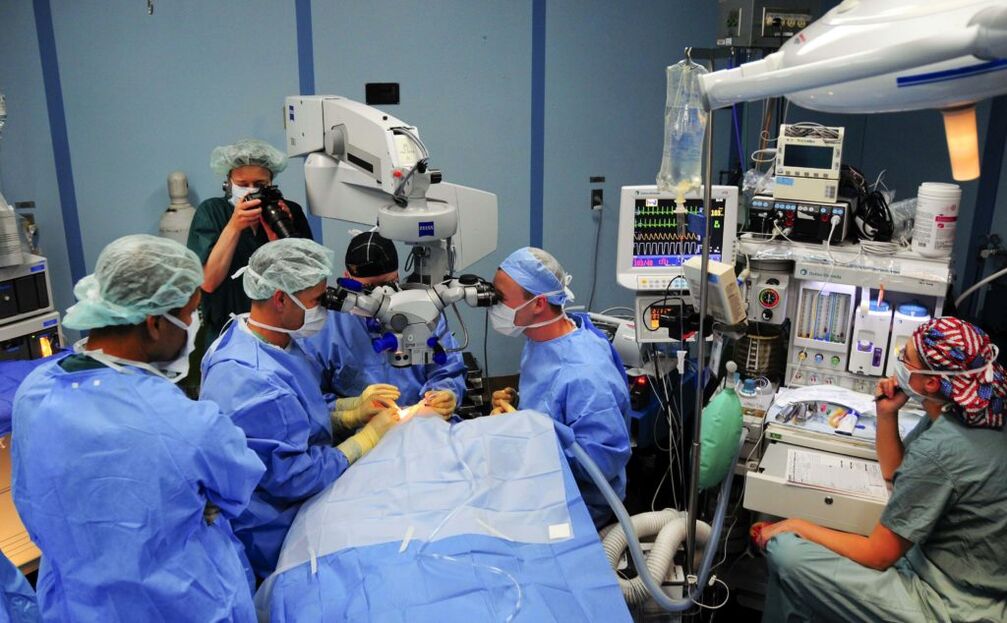
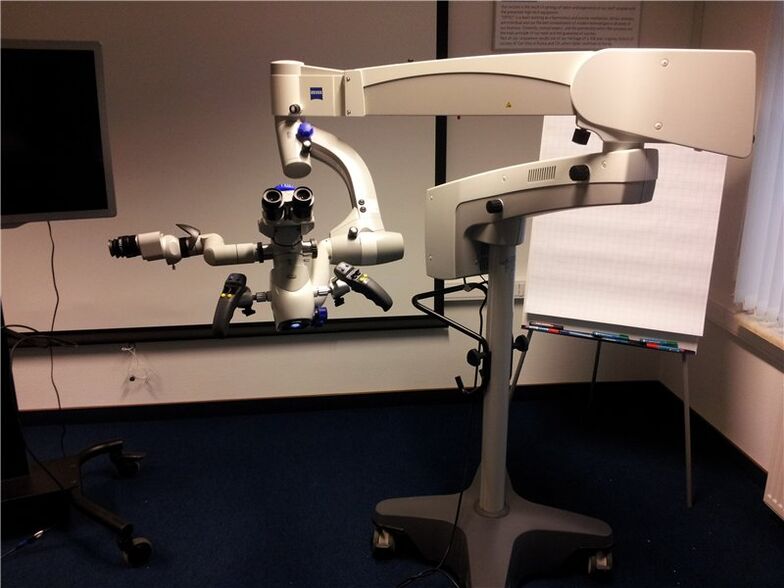
Before the operation, it is necessary to undergo diagnostics, the doctor will voice the list of tests at the consultation.Methods of correction of vision are selected individually by every patient, they can be recommended to the patient with cataracts, myopia, strabismus, halazion, glaucoma and other dangerous deviations.During manipulations, ophthalmologists use modern medical equipment, intervention is carried out using a microscope, vision is adjusted through very small incisions.Since the operation is simple and low -traumatic, long -term hospitalization is not required.After 2-3 hours, the doctor explains in detail to the patient all the nuances of the recovery period, how to care for the eye, and lets go home.The hospital is recommended only with serious pathologies (retinal detachment, severe inflammations, penetrating wounds).There are such types of operations:
- laser surgery;
- vitrectomy;
- Trabekulectomy;
- LTP, Lie;
- focoemulsification;
- scleroplasty;
- keratoplasty;
- Enucleation of the eye;
- refractive replacement of the lens;
- Crosslinking;
- anti -hacomatous operations.
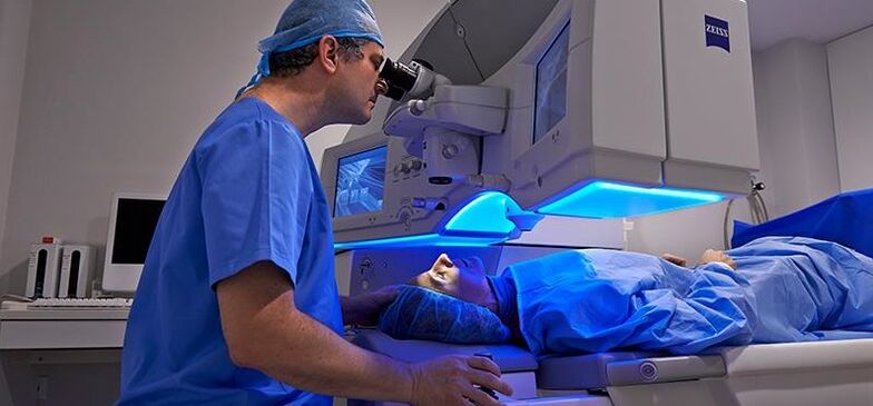
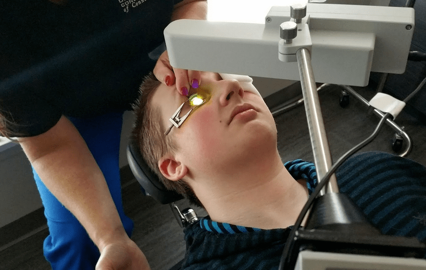
Laser vision correction
Do the operation for damage and ruptures of the retina.Before agreeing, the patient must undergo a thorough examination.Drops that expand the pupil are dripping before the intervention.With the help of a laser, the eyes are made by microsurgery (weat weak areas of tissues are soldered), this prevents the penetration of fluid, and subsequently detachment.Laser coagulation of the retina is simple, takes several minutes in time, even kids tolerate it well.
The operation takes place under local anesthesia.In the recovery period, the patient is recommended to abandon active sports, bad habits, and physical overloads.The negative consequences of laser vision correction can manifest itself within 3 hours - this is an increased secretion of tears, photophobia, redness, discomfort and rub in the eye.Doctors do not recommend this procedure under 18 years of age.The percentage of vision restoration after laser correction is very large.
Keratoplasty
With irreversible pathological changes in the eye, the Okulists recommend this procedure.The operation to restore vision and eyes after the injury consists in the complete removal of pathologically altered fabrics of the cornea, which are then replaced by donor.There can be many reasons for surgical intervention - it is keratoconus, severe damage to the tissues of the eyeball, EED, ulcerative changes caused by fungal infections, and genetic predisposition.In surgery, local anesthesia is used.Using the instruments, the doctor needs to remove the sore cornea and replace it with a healthy one, after which the seams are applied.The time takes about 2 hours.The recovery period is 1 year.Eyes are recommended to protect with glasses, this will prevent injury to the organ, complications after surgery.
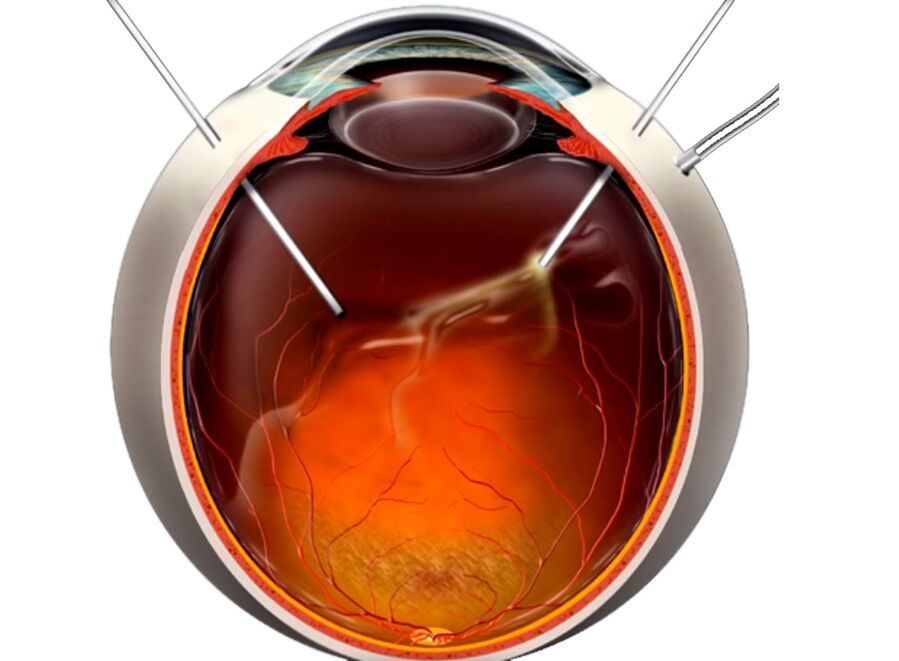
Vitrectomy
Make vision correction with pathologically altered vessels on the retina and when it is removed.Thanks to professionally conducted manipulations, the doctor will gain access to the rear side of the eye.After removal, the vitreous body is replaced by donor material.A saline solution, silicone oil, polymers and perfactor -urgent compounds are used as a special liquid.In ophthalmology, vitrectomy is considered a micro -vasic methodology, interference in the system of the visual organ is minimal, this contributes to the patient's return to the patient.With the help of this method, it is possible to restore the structure of the retina, to prevent the development of pathologies, the names of which are traction and retinopathy.
Since the air is filled with many dust particles, in order to prevent infection after the organ correction, it is necessary to tightly glue the operated eye with a bandage for some time.
Anti -hacomatous operations
Eye surgery should be used if treatment with drugs is unauthorizing, and only after a complete examination.This type of intervention does not cause pain, it is made quickly, usually prescribed for the treatment of closed -angled and open -angle glaucoma, and when the vessels germinate inside the eyes.The essence of the procedure is to eliminate the accumulated fluid inside the organ, it is carried out by a laser.The most effective methodology is considered non -praying deep sclerectomy.In the process of therapy, the corneal layer surgically interpret, thereby reducing increased intraocular pressure.Possible consequences of operations of this kind can be expressed in the formation of scars, at first the eyes can be red.
Crosslinking
A fairly new method of treating keratoconus, using it, can prevent the development of the disease in the initial stages.Using a tool whose name is a boring lamp, and dripping vitamin B into the eye socket2, it is possible to strengthen the cornea and stabilize the patient's condition.The plus of the procedure is that manipulation is carried out only once, 1 hour is enough to carry out it.After to reduce the risks of injury, a special protective lens is put on the eyeball.
Enucleation of the eye
This is a complex surgical operation, it is prescribed only in extreme cases, if an organ cannot be saved in a different way.The eyes are completely removed from the eye socket, and the fiberglass prosthesis is implanted in its place, it will look like real.When conducting, side effects may develop, these are unforeseen inflammatory processes and implant displacement.The testimony includes such pathologies as:
- glaucoma to a terminal degree;
- the appearance of pain and the development of inflammation in the blind organ;
- Serious injuries and neoplasms.
Refractal replacement of the lens
During surgery, the damaged element of the visual system with an artificial system occurs.When selecting an intraocular lens, the individual characteristics of the patient are taken into account (age, gender).The procedure lasts 25 minutes, local anesthesia is used, surgical manipulations take place without blood.Through microdnaras, using ultrasound, the lens is turned into an emulsion and removed from the eye.Refraction replacement is made with pathologies such as:
- running myopia and hyperopy;
- presbyopia;
- prohibition on laser correction;
- High -degree hyperopy, which can cause glaucoma;
- Violation of refraction and quick loss of visual function.
Eximerlazer vision correction
Surgical intervention is recommended for eye pathologies such as strabismus, astigmatism, farsightedness.There are several ways to carry out, this is a femo lasik, Federal Republic of Criminal Code, Lasik.The doctor cuts off the upper layer of the cornea, its curvature is changed using a cold beam of an excessive laser, it is better to use local anesthesia and a mediocre.After an eximerlazer correction, the patient gets rid of the problem, since the procedure is effective by 99%, in the plus in front of other interventions.An unsuccessful operation leads to hyper -correction, causes redness, and the corneal layer looks inflamed.
Correction of strabismus
Eye surgery for strabismus is aimed at correcting the symmetrical position of the organ.Surgical intervention is divided into two types: this is an increase in the muscles that are responsible for the movement of the visual organ and the relief in the oculomotor muscle.In the first case, a part of the muscle system, as well as anti -position and tenorafia, is resected, in the second they change the place of muscle fastening, lengthen it by plasticity.What method to turn to, the surgeon decides, given the individual factors and the degree of severity of the pathology.
Scleroplasty
Doctors recommend referring to this type of intervention with uncontrolled development of myopia.Using the methodology, strengthen and stabilize the patient's condition, stop loss of vision.Scleroplasty is shown to children and adolescents at the age of 17 during the active period of growth, with such pathologies of the eye as rapidly developing myopia.
After removing the eye in the early days, the patient may feel severe pain, in this case, you need to consult your doctor and choose the right analgesics.
Removal of formations
Surgical intervention of this type is indicated for halazium, cystic neoplasms of conjunctiva and ptergium.Holding the growth with tweezers, the doctor cuts it out further to eliminate negative consequences, the wound is cauterized.To prevent infection, an antibiotic ointment is applied over the eyelids, and a sterile bandage is used.The disadvantage is that the disease can develop again, in this case, return to this method of therapy cannot be avoided.
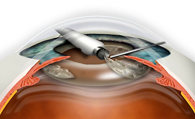
Removal of cataracts
During the microsurgery, a pathologically changed lens is removed, therapy is carried out using laser and ultrasonic phacoemulsification.Special surgical tools through micronads are thinned out and removed from the eye, replaced with artificial.Sometimes they do a procedure called intracapsular and extracapsular extraction.